
The system
How QuietScan works
The child sits on a parent’s lap and wears a soft cap. The session is designed to last under five minutes, with no sedation. The clinician receives a 3D map of active and quieter regions — a monitoring view, not a diagnosis and not a treatment.
Physics and design follow. MATLAB and ANSYS cases are simulations. The wearable has not been fabricated.
- Intended for infants and children ages 0–5
- Designed for no sedation
- Intended session under 5 minutes
- Intended for use on a parent’s lap
- 64 transducers in eight concentric groups
- Skull-aberration correction and AI delay estimation (in development)
- Form factor
- Wearable cap / helmet — in development
- Array
- 64 piezoelectric elements, 8 concentric groups
- Session
- Intended: under 5 minutes, no sedation, parent lap
- Software
- AI delay estimation + 3D map — in development
- Calibration
- Optional genetic markers — roadmap, not a current feature
- Regulatory
- Intended FDA 510(k) Class II diagnostic ultrasound
Lab trials
How the beam finds the brain
Working simulation cases. Not human clinical data. Not a diagnostic output.
Green is energy in the right place. Red is energy that missed.
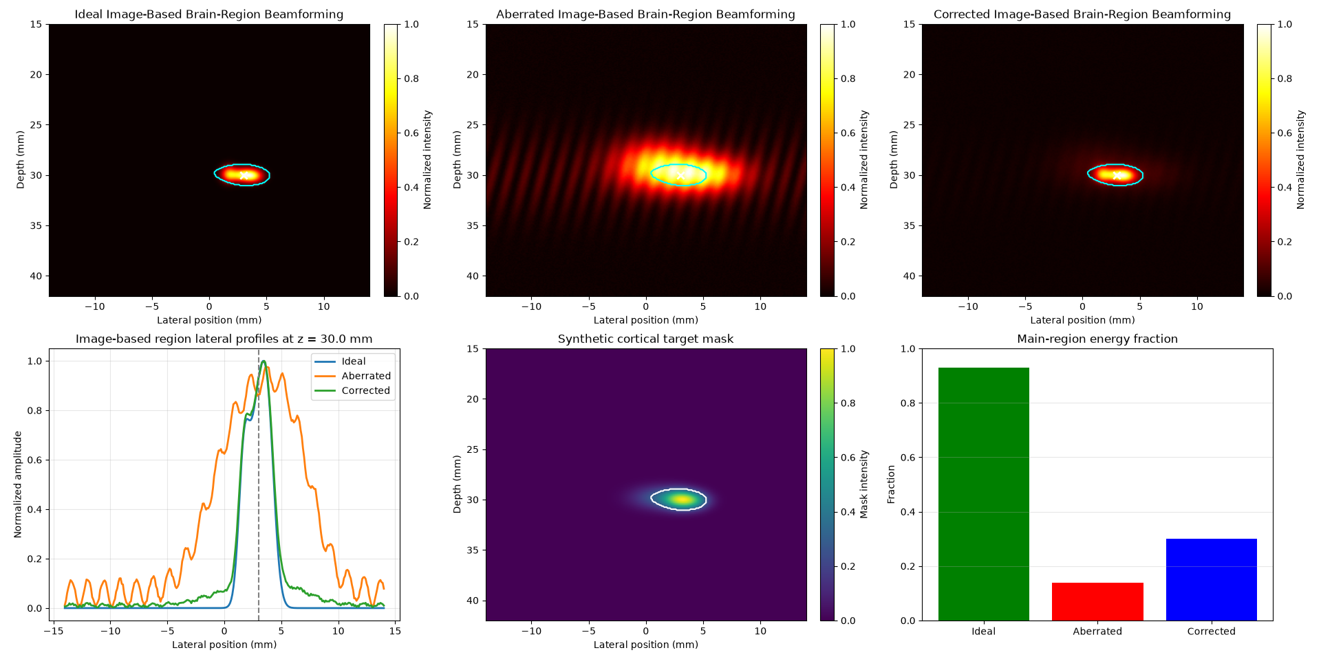
Case 4 — Image-based cortical target
Image-based focusing on a synthetic cortical mask. After the skull, the beam should stay in the region — not smear into neighboring tissue.
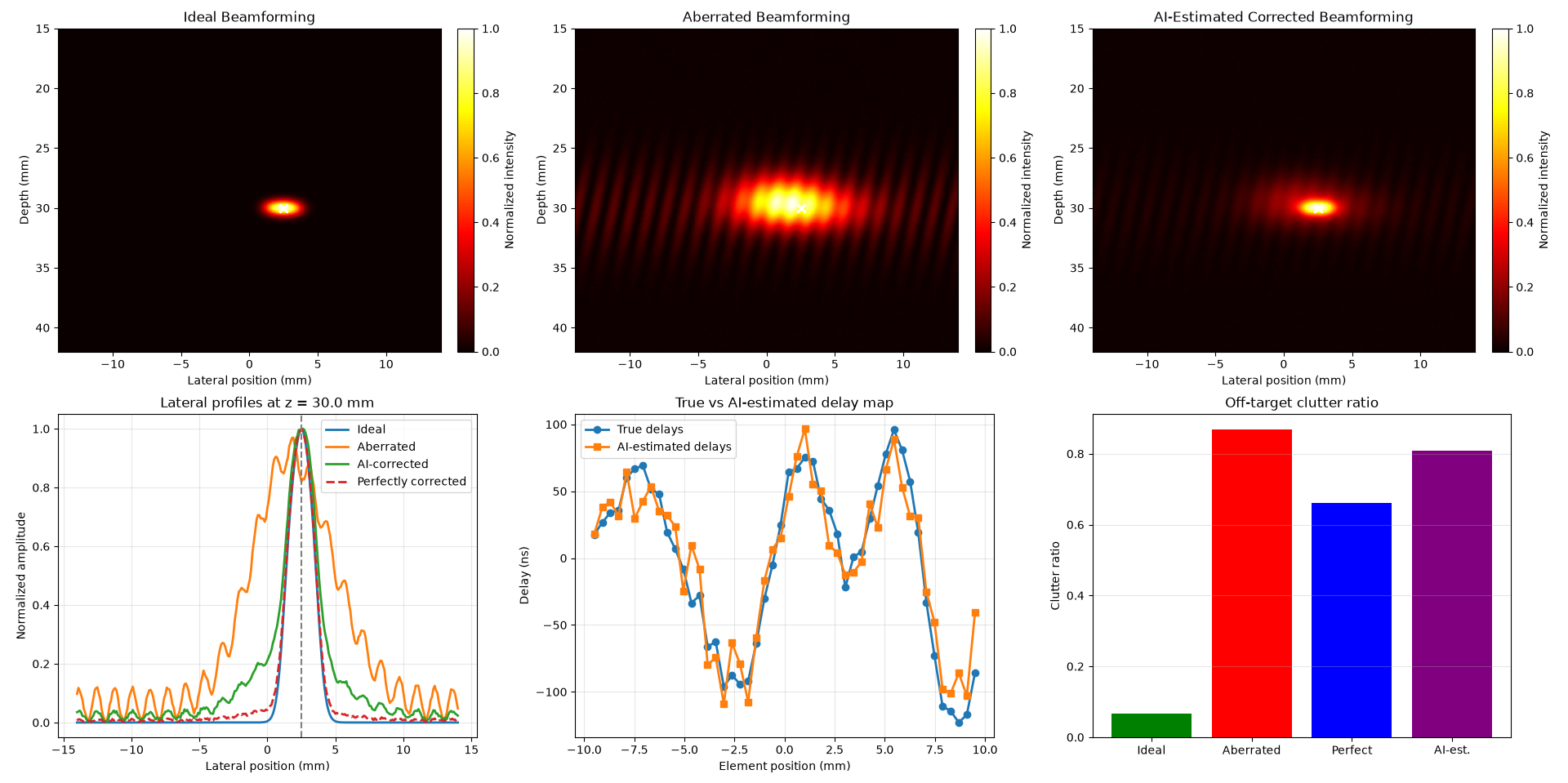
Case 5 — AI-estimated delays
An AI delay map is estimated element by element and compared with the true delays. AI-corrected focus approaches perfect correction; clutter falls from the aberrated case toward the ideal.